Coronary stenosis: grading, reference diameter and total occlusion
David F. Cuevas Cueto, MD · Updated September 27, 2026 · Sources: SCCT 2026 and CAD-RADS 2.0
Stenosis is graded by the reduction in luminal diameter relative to a normal reference segment and is reported as a term with its range. The patient's CAD-RADS category is based on the most severe stenosis, a left main stenosis of 50% or more, or stenosis of 70% or more in all three vessels, raises it to 4B. Definitions follow the SCCT 2026 consensus and CAD-RADS 2.0.
Key points
Stenosis is graded by diameter, comparing the minimal lumen with the lumen of a nearby normal segment, never with the outer contour of a remodeled plaque.
The grades are minimal (1–24%), mild (25–49%), moderate (50–69%), severe (70–99%) and total occlusion (100%), and the report always gives the term with its range.
A visual estimate of the stenosis range is sufficient for the clinical report; quantitative measurement is not a routine requirement and varies by about ±25% compared with invasive angiography.
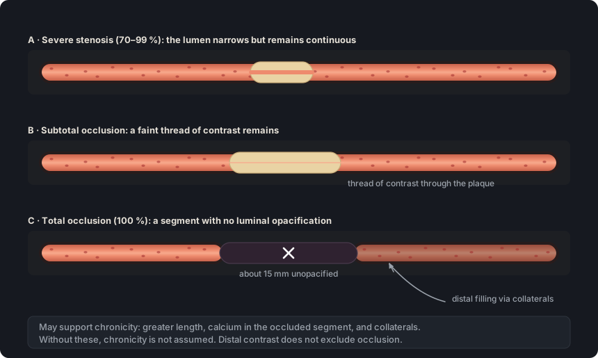
A segment with no luminal opacification is a total occlusion even when there is distal contrast, and chronicity is not assumed but inferred from collaterals, calcium or length.
A stenosis is obstructive at 50% or more and severe at 70% or more, but in the left main a stenosis of 50% or more is already CAD-RADS 4B.
Each stenosis is described by segment, with its anatomical name, plaque type and grade; in serial or diffuse disease, the most severe stenosis sets the category.
Four segments were marked with buttons 1 to 4. The template writes each grade with its term and range, and the severe mid RCA stenosis sets the category (CAD-RADS 4A/P2).
Table 1. Coronary stenosis grading scale by diameter. The category shown applies to the patient when that grade is the most severe stenosis; a left main stenosis of 50% or more is CAD-RADS 4B.
Grade
Term
Range
Meaning
0
No visible stenosis
0%
No luminal narrowing. Without plaque, CAD-RADS 0; with plaque, CAD-RADS 1
1
Minimal
1–24%
Nonobstructive (CAD-RADS 1)
2
Mild
25–49%
Nonobstructive (CAD-RADS 2)
3
Moderate
50–69%
Obstructive (CAD-RADS 3; 4B if in the left main)
4
Severe
70–99%
Obstructive (CAD-RADS 4A; 4B if in the left main or in three vessels)
5
Total occlusion
100%
Nonopacified segment, acute or chronic (CAD-RADS 5)
Source: SCCT 2026, text p. 7 and Table 5, p. 20; CAD-RADS 2.0, Tables 1 and 4. Table 5 gives “minimal (1–25%)”; the SCCT 2026 text and CAD-RADS 2.0 give “1–24%”.
Also seen as:percent diameter stenosis, DS, luminal diameter reduction
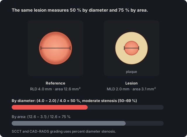
Diameter stenosis is the percent reduction in luminal diameter at the narrowest point relative to a normal reference segment. SCCT and CAD-RADS grade stenosis by diameter, not by area.
Do not confuse: diameter stenosis with area stenosis. In a circular lumen, 50% diameter stenosis equals 75% area stenosis.
Source: SCCT 2026, qualitative stenosis grading, p. 7; curved MPR, p. 2; formula under quantitative grading, p. 7 · “A qualitative interpretation of the extent of coronary artery stenosis should be made in transverse and longitudinal views as an estimated maximal percent diameter stenosis.”
Details
In practice. Maximal stenosis is estimated in transverse and longitudinal views, because curved MPR can underestimate an eccentric stenosis. If measured, it is calculated as (RLD − MLD) / RLD × 100, where RLD is the reference luminal diameter and MLD the minimal luminal diameter.
Pitfall. Reporting percent area stenosis as if it were diameter stenosis. Area gives higher figures for the same lesion, and the CAD-RADS scale applies to diameter.
Other sources. SCCT 2014 treated area stenosis as optional additional information, not a required element. (SCCT 2014, section 5.4, p. 346)
In CCTA Map
The 0–5 buttons and the “Stenosis grade” slider record the percentage on the CAD-RADS diameter scale, and each segment takes its highest-grade lesion. There is no field for area stenosis.
Figure 1. The same lesion measures 50% by diameter and 75% by area, because area is proportional to the square of the diameter. The SCCT and CAD-RADS scale applies to percent diameter stenosis.
Also seen as:reference diameter, reference lumen diameter, reference vessel diameter(quantitative coronary angiography term, where it measures the lumen), RVD(quantitative coronary angiography abbreviation)
The reference luminal diameter is measured in the lumen of a normal segment near the stenosis, proximal or distal, or taken as the average of both. It is the denominator of percent stenosis.
Do not confuse: the reference luminal diameter with the outer vessel diameter. The outer diameter is used to assess positive remodeling; stenosis is calculated from luminal diameters only.
Source: SCCT 2026, qualitative (p. 7) and quantitative (p. 7) stenosis grading; overestimation, p. 17 · “Qualitatively, the luminal diameter at a stenosis should be compared with the luminal diameter of a normal reference vessel segment distal and/or proximal to the stenosis.”
Details
In practice. Choose the most normal cross-section within 10 mm of the lesion, with no branch vessels in between. For an ostial lesion, use the nearest distal reference. In the left main and in diffuse disease, normal reference diameters can be used.
Pitfall. Comparing the lumen with the outer contour of a positively remodeled plaque overestimates stenosis. The minimal lumen is compared with the lumen of a normal segment.
Other sources. The 2024 ASCI-PT consensus prefers the most normal-appearing site immediately proximal to the plaque. If the proximal segment is unsuitable, as in an ostial lesion, it uses the nearest distal reference. An interpolated reference remains an option for comparison with quantitative coronary angiography. (Kim 2024, ASCI-PT, topic 7 and Table 4)
In CCTA Map
The map does not measure diameters. The percentage recorded with the buttons or the slider is the one the radiologist estimated or measured at the workstation.
Also seen as:coronary stenosis grades, stenosis grading, critical stenosis(not part of the scale; write the grade with its range)avoided in this glossary
SCCT 2026 and CAD-RADS 2.0 divide diameter stenosis into six grades: 0%, minimal (1–24%), mild (25–49%), moderate (50–69%), severe (70–99%) and total occlusion (100%).
How to write it
Proximal LCx: calcified plaque with minimal stenosis (1–24%).
Do not confuse: the stenosis grade with the plaque burden. CAD-RADS 2.0 uses “mild”, “moderate” and “severe” on both scales, and a minimal stenosis can accompany severe plaque burden (CAD-RADS 1/P3).
Source: SCCT 2026, qualitative stenosis grading, p. 7 · “This stenosis can be categorized as minimal (1-24 %), mild (25-49 %), moderate (50-69 %), severe (70-99 %) and total occlusion (100 %)”
Details
In practice. In the report, the term is written with its range, for example “moderate stenosis (50–69%)”, so that it is compatible with invasive angiography categories. The CAD-RADS category is based on the patient's most severe stenosis.
Pitfall. Writing only “moderate stenosis” without the range, or using words outside the scale, such as “critical”. Write a term from the scale followed by its range.
Other sources. SCCT 2009 allowed an optional scale in which mild was less than 39% and moderate 40 to 69%. Some studies also use other cutoffs for mild and minimal. Before comparing an article with this scale, check which cutoffs it used. (Raff 2009, SCCT, optional quantitative scale; Kim 2024, ASCI-PT, topic 6 and Table 3)
In CCTA Map
The editor's 0–5 buttons store a representative value for each range (0, 15, 38, 60, 85 or 100%), and the template writes the term with its range, for example “moderate stenosis (50-69%)”. That internal value is not a measurement: the editor shows it next to the “Stenosis grade” slider, but the report does not print it.
Literal output of the map's template
Left anterior descending (LAD): proximal segment: calcified plaque causing minimal stenosis (1-24%); mid segment: partially calcified plaque resulting in mild stenosis (25-49%); the remainder is unremarkable. Branches D1, D2: no plaque.
Circumflex (LCx): proximal segment: non-calcified plaque causing moderate stenosis (50-69%); the rest of the vessel is unremarkable. Branches OM1, OM2, L-PLB: no plaque.
Right coronary (RCA): mid segment: partially calcified plaque producing severe stenosis (70-99%); the rest of the vessel is unremarkable. Branches AM, R-PDA, R-PLB: no plaque.
Impression Severe obstructive CAD. CAD-RADS 4A/P2.
Also seen as:total coronary occlusion, complete occlusion, 100% stenosis, occluded segment, chronic total occlusion, CTO(only with findings that support chronicity, such as collaterals; CAD-RADS 5 does not imply it), subtotal occlusion(not a total occlusion: it retains minimal luminal opacification), collateral circulation, collaterals(collaterals fill the bed distal to the occlusion and support its chronicity)
A total occlusion is a coronary segment with no luminal opacification, equivalent to 100% stenosis. A single total occlusion, acute or chronic, makes the study CAD-RADS 5.
How to write it
Mid RCA: total occlusion (100%) with opacification of the distal RCA.
Do not confuse: total occlusion with subtotal occlusion. A subtotal occlusion retains minimal luminal opacification, which can be difficult to detect, and is generally more amenable to intervention than a chronic total occlusion.
Source: SCCT 2026, coronary occlusion (total, subtotal and features of chronicity), p. 10 · “Total coronary occlusion is defined by complete nonopacification of a coronary artery segment”
Details
In practice. Follow the segment on curved MPR and transverse views. If a thread of contrast crosses the lesion, it is a subtotal occlusion. Distal contrast does not exclude occlusion, because the delayed acquisition allows time for collateral filling.
Pitfall. Writing “chronic occlusion” or “CTO” only because the segment is occluded. Chronicity is inferred separately, from findings such as collaterals, calcium or greater length.
Other sources. In the catheterization laboratory, chronic total occlusion requires absent antegrade flow through the lesion and a probable or documented duration of three months or more. On CCTA, length, calcium and collaterals only suggest chronicity. (Galassi 2024, EAPCI consensus on chronic total occlusions, introduction; SCCT 2026, p. 10; SCCT 2024 quantification, Table 4, p. 437)
In CCTA Map
The “5” button in the vessel editor sets the lesion to 100%, the template writes “total occlusion (100%)” and the category becomes CAD-RADS 5. The template adds “consistent with chronic total occlusion” only when a collateral that reaches the occluded territory is turned on in the “Collateral circulation” panel of the “Anatomy ▾” menu.
Literal output of the map's template
Right coronary (RCA): mid segment: non-calcified plaque, total occlusion (100%); the rest of the vessel is unremarkable. Branches AM, R-PDA, R-PLB: no plaque.
Distal reconstitution of the occluded RCA via collaterals (from the LAD · apical PDA–distal LAD anastomosis), consistent with chronic total occlusion.
Impression Total occlusion (100%) of at least one vessel. CAD-RADS 5/P1.
Obstructive CAD (≥50%) in RCA.
The occlusion demonstrates collateral recruitment to the distal territory, supporting chronicity (chronic total occlusion) and relevant to planning the revascularization strategy and access route (e.g., CTO-PCI). Confirm myocardial viability with dedicated imaging, as collaterals alone do not reliably predict it.
Figure 2. Total occlusion is defined by absent opacification of a segment, and distal contrast does not exclude it because it can arrive through collaterals. A thread of contrast through the lesion indicates subtotal occlusion.
Also seen as:patency, no visible stenosis(CAD-RADS 2.0 term for 0%), no luminal narrowing, no plaque or stenosis, no atherosclerotic disease(equivalent to no plaque), no significant stenosis(ambiguous: it does not say whether there is plaque or which cutoff is used)avoided in this glossary, normal, unremarkable(only if there is no plaque or stenosis)
These are three different statements. “Patent” means that the lumen of a vessel, stent or graft opacifies with contrast and is not occluded, but it does not say whether the lumen is narrowed. “No stenosis” means that the lumen is not narrowed (0%), although plaque may be present. “No plaque” means that the wall has no plaque. A segment is normal only if it has no plaque and no stenosis.
How to write it
Mid RCA: calcified plaque without visible luminal narrowing.
Do not confuse: “patent” with “no stenosis”. A patent stent can have moderate restenosis (50–69%), and a patent graft can have a stenosis at the distal anastomosis.
Source: SCCT 2026, coronary stent (p. 10), grafts (pp. 11–12), atherosclerotic plaque (p. 5) and impression (p. 21); CAD-RADS 2.0, Table 1 (0%, “No visible stenosis”) and Table 4 (CAD-RADS 0 and 1, note b) · “An impression of a normal study should be given only if there is no plaque or stenosis.”
Details
In practice. In a stent, patency is stated when there is contrast within the stent; distal contrast alone is not enough. In a graft, report patency and, separately, any stenosis of the graft body, the anastomoses and the distal runoff. In a segment with plaque that does not narrow the lumen, write the plaque type followed by “without visible luminal narrowing”. Before writing “no plaque”, review the wall on cross-sectional views: a small, low-attenuation calcified plaque can be isoattenuating to the contrast-filled lumen, and the non-contrast scan helps to detect it.
Pitfall. Writing “patent coronary arteries” or “no significant stenosis” as if it meant a normal study. SCCT 2026 reserves the impression of a normal study for the absence of plaque and stenosis, and plaque that does not narrow the lumen already means at least CAD-RADS 1.
In CCTA Map
By default, the map uses the normal template: unmarked segments are written with the per-vessel wording (“no atherosclerotic disease”, “no plaque or stenosis”, “the remainder is unremarkable”), and a gray line under the Word and PDF buttons says so (“Anything you don't mark is described as normal. Review it before use.”); the line is not carried into the copied text, the Word file, the printed sheet or the PNG. With “nondiagnostic” under “Quality”, that wording becomes “no identifiable lesions”, and with P set by “Calcium (Agatston)” or “Pick P” and no plaque drawn, “no stenosis”. The “Not evaluated” button in the editor records a segment that was not assessed. A stent without restenosis is written as “patent stent, no significant restenosis”, and a graft without stenosis as “patent, no significant stenosis”. A plaque at 0% is written with its type, as in “non-calcified plaque without significant stenosis”.
Obstructive and nonobstructive stenosis ≥50% · ≥70%#
Also seen as:obstructive CAD, nonobstructive CAD, obstructive coronary artery disease, nonobstructive coronary artery disease, significant stenosis(ambiguous: depending on the source it means 50% or more, 70% or more, or a functional effect; the grade with its range is preferred)avoided in this glossary
In CAD-RADS 2.0, which this glossary follows, a stenosis of 50% or more is called obstructive, and one below 50% nonobstructive. The 70% cutoff marks severe stenosis and defines obstructive three-vessel disease.
How to write it
Obstructive coronary artery disease, with a maximal stenosis of moderate grade (50–69%) in the mid LAD.
Do not confuse: “obstructive” with “hemodynamically significant”. “Obstructive” is an anatomic threshold, a diameter stenosis of 50% or more; hemodynamic significance is assessed with a functional test and, if that test was CT-FFR or CT perfusion, its result is coded with the I modifier.
Source: CAD-RADS 2.0, section 3.1.1 and Table 4 (CAD-RADS 1 and 2 as nonobstructive disease; interpretation of CAD-RADS 3 to 4B) · “CAD RADS 4B - This indicates the presence of a left main stenosis of at least 50% or three-vessel obstructive disease (>70%).”
Details
In practice. The 50% cutoff separates CAD-RADS 2 from CAD-RADS 3, for which CAD-RADS 2.0 suggests considering functional testing. The 70% cutoff separates CAD-RADS 3 from 4A. A left main stenosis of 50% or more, or three-vessel disease with 70% or more in each vessel, is CAD-RADS 4B.
Pitfall. Classifying a 50 to 69% left main stenosis as CAD-RADS 3. In the left main, a stenosis of 50% or more is CAD-RADS 4B.
Other sources. The 2021 ACC/AHA/SCAI revascularization guideline defines a significant angiographic stenosis as 70% or more, or 50% or more in the left main, and an intermediate stenosis as 40–69%. The glossary follows the 50% cutoff of CAD-RADS 2.0. (Lawton 2022, ACC/AHA/SCAI revascularization guideline, section 4.1)
In CCTA Map
The impression calls CAD-RADS 1 and 2 “nonobstructive” and lists, by abbreviation, where the obstructive disease (≥50%) is: the vessel (LM, LAD, LCx or RCA) if the lesion is in it, and the branch (D1, OM1, R-PDA or RI, for example) if it is in a branch, as in “Obstructive CAD (≥50%) in LAD, D1 and R-PDA”. The nonobstructive disease line names them the same way, for example “Nonobstructive CAD in D1 and OM1”. A left main stenosis of 50% or more gives CAD-RADS 4B even without a severe stenosis. Only atherosclerotic stenosis counts: a narrowing marked “Non-atherosclerotic” does not give 4B and is not included in these lists; the impression names it separately, with the E modifier.
Literal output of the map's template
Left anterior descending (LAD): proximal segment: partially calcified plaque causing moderate stenosis (50-69%); the remainder is unremarkable. Branches D1, D2: no plaque.
Circumflex (LCx): proximal segment: partially calcified plaque causing moderate stenosis (50-69%); the rest of the vessel is unremarkable. Branches OM1, OM2, L-PLB: no plaque.
Right coronary (RCA): mid segment: partially calcified plaque producing moderate stenosis (50-69%); the rest of the vessel is unremarkable. Branches AM, R-PDA, R-PLB: no plaque.
Also seen as:per-lesion analysis, per-segment analysis, per-vessel analysis
These are the levels at which coronary disease is described. A lesion is a specific plaque, with its extent and its maximal stenosis, and it can involve more than one segment. A segment is the unit of the SCCT segment model, in which plaque type and stenosis are recorded. A vessel is the artery made up of those segments. The CAD-RADS category is assigned to the patient, not to each lesion or each vessel.
How to write it
Partially calcified plaque extending from the proximal to the mid LAD, with a maximal stenosis of moderate grade (50–69%) in the mid LAD.
Do not confuse: the lesion with the segment when counting the SIS. A plaque that involves the proximal and mid LAD is a single lesion but adds 2 points; three plaques in the same segment add 1.
Source: SCCT 2026, atherosclerotic plaque and plaque volume (p. 5) and Table 6 (per-segment assessment of plaque and stenosis), p. 20; CAD-RADS 2.0, sections 3.1.1 and 3.3.4, Table 4 (note on per-patient classification) and Figure 17 · “Depending on the extent of disease, plaque can be described on a per-lesion or per-segment level.”
Details
In practice. Maximal stenosis and high-risk features belong to a lesion, and HRP requires two or more features in the same plaque. The report records plaque type and stenosis per segment, and the SIS counts segments with plaque, not plaques. Three-vessel disease for CAD-RADS 4B is counted per vessel. The patient's category is set by the clinically most relevant stenosis, usually the highest-grade one. SCCT 2026 allows plaque to be described per lesion or per segment, depending on the extent of disease.
Pitfall. Recording a long plaque only in the segment of its maximal stenosis. The plaque is recorded in every segment it involves, and each one adds to the SIS.
In CCTA Map
Each lesion is drawn by dragging along the vessel and is characterized in the window that opens. A plaque dragged across two segments is a single lesion, with one plaque type and one set of features, and counts toward the SIS in both; each stretch has its own grade, by default that of the lesion, which can be lowered in the editor, and the template describes it once, in the most proximal segment. High-risk features (“Low attenuation”, “Positive remodeling”, “Spotty calcification” and “Napkin-ring sign”) are marked on each plaque, and HRP appears only when a single plaque has two or more. If a segment has several plaques, the template names them all, from the highest grade down (“in the same segment, another…”), each with its type, grade and features; a stent and a native plaque in the same segment are also described separately. The app shows a single per-patient category with its reason, for example “Three-vessel disease (≥70%)”.
Also seen as:serial lesions, serial stenoses, sequential stenoses, tandem stenoses, tandem lesions(in SYNTAX, lesions count as tandem only when separated by less than three reference diameters), diffuse coronary artery disease, diffuse plaque, multifocal disease(SCCT 2026 attribute “multifocal”), focal lesion, focal stenosis, long lesion(SCCT 2026 attribute “long”)
These terms describe the extent of disease along the vessel. A focal lesion involves a short length, with normal wall on both sides. Serial stenoses are two or more stenoses in the same vessel separated by normal lumen; “multifocal” describes several lesions separate from one another. In diffuse disease, plaque extends over a long length with no normal segment nearby. SCCT 2026 does not set a length that separates focal from diffuse atherosclerotic lesions.
How to write it
Mid LAD: two non-calcified plaques in series, the proximal one with mild stenosis (25–49%) and the distal one with severe stenosis (70–99%).
Do not confuse: serial stenoses with diffuse disease. In serial stenoses, normal lumen remains between the stenoses and each one is graded separately; in diffuse disease, there is no nearby normal lumen to serve as a reference.
Source: SCCT 2026, atherosclerotic plaque (p. 5), quantitative grading (p. 7) and underestimation (p. 18) · “Additional attributes such as "ostial", "bifurcation", "eccentric", "long", "diffuse" and "multifocal" can be used to further describe the location, morphology and extent of atherosclerotic disease.”
Details
In practice. SCCT 2026 allows plaque to be described per lesion or per segment, with attributes such as “long”, “diffuse” or “multifocal”. In diffuse disease, name the portion of the vessel that the plaque involves and the site of maximal stenosis; in serial stenoses, describe each one with its grade. The CAD-RADS category is based on the most severe stenosis, and extent is reflected in plaque burden, because every segment with plaque adds to the SIS.
Pitfall. In diffuse disease, using a neighboring segment that is also diseased or small as the reference underestimates stenosis. Use the expected normal diameter of the vessel instead.
Other sources. In the SYNTAX score, lesions separated by less than three reference diameters count as a single tandem lesion. Its diffuse disease criterion describes not plaque but a distal vessel smaller than 2 mm over at least 75% of its length. SCCT 2026 gives lengths for “focal” and “diffuse” only within specific classifications, in-stent restenosis (10 mm) and spontaneous coronary artery dissection (20 mm), and does not apply them to atherosclerotic plaque. (Sianos 2005, SYNTAX, pp. 222–223; SCCT 2026, pp. 10 and 13)
In CCTA Map
If two lesions are drawn in the same segment, the template first describes the highest-grade one with its plaque type and then the other, in a clause of its own (“in the same segment, another…”); the category is based on the most severe stenosis. A plaque dragged across two segments is a single lesion that counts toward the SIS in both; the template describes it once, with its extent. The map has no field for “focal”, “diffuse” or “multifocal”; write these attributes in the draft.
Literal output of the map's template
Left anterior descending (LAD): proximal segment: non-calcified plaque causing mild stenosis (25-49%); mid segment: non-calcified plaque resulting in severe stenosis (70-99%); in the same segment, another non-calcified plaque resulting in mild stenosis (25-49%); the remainder is unremarkable. Branches D1, D2: no plaque.
Impression Severe obstructive CAD. CAD-RADS 4A/P1.
Also seen as:eccentric plaque, eccentric stenosis, concentric plaque, circumferential plaque
These terms describe how plaque is distributed around the circumference of the wall. Eccentric plaque involves only part of the circumference, and the lumen is displaced toward the uninvolved side. Concentric plaque surrounds the lumen along its entire circumference. Neither word indicates the degree of stenosis.
How to write it
Mid RCA: eccentric non-calcified plaque with mild stenosis (25–49%).
Do not confuse: concentric plaque with the smooth, circumferential wall thickening of vasculitis, which enhances with contrast and can be accompanied by stranding of the adjacent fat. This thickening is not atherosclerotic and is coded with modifier E.
Source: SCCT 2026, atherosclerotic plaque (“eccentric” attribute, p. 5), curved MPR (p. 2) and vasculitis (p. 13); CAD-RADS 2.0, section 3.3.6; Kim 2024, ASCI-PT, topic 8 · “Curved MPR can underestimate eccentric stenosis; hence evaluation should be performed in at least two perpendicular planes.”
Details
In practice. Curved MPR can underestimate an eccentric stenosis, so stenosis is assessed in at least two perpendicular planes and on cross-sectional views. If an elliptical lumen is measured on a cross-sectional view, ASCI-PT 2024 advises averaging the maximal and minimal diameters, with moderate agreement (66.7%).
Pitfall. Grading an eccentric plaque in a single longitudinal plane. The lumen can look wide in that plane and narrow in the perpendicular one.
Other sources. SCCT 2014 listed eccentric position among the optional descriptors of a stenotic lesion. In SCCT 2026, “concentric” appears in cardiac allograft vasculopathy, which causes distal concentric narrowing; in these patients, proximal eccentric plaque can be due to donor atherosclerosis. (SCCT 2014, section 10.6.2, p. 352; SCCT 2026, p. 13)
In CCTA Map
The map does not record whether plaque is eccentric or concentric. Add the attribute when editing the draft.
Also seen as:visual estimation of stenosis, stenosis quantification, visual assessment, eyeballing(colloquial)
In visual estimation, the reader assigns the stenosis range by comparing the lumen with the reference by eye. Quantitative measurement calculates the percentage from diameters or areas traced by software.
How to write it
Mid LAD: partially calcified plaque with moderate stenosis (50–69%), visually estimated at approximately 55%.
Do not confuse: a visually estimated figure with a measurement. If no diameters were traced, present the figure as a visual estimate, together with the term and its range.
Source: SCCT 2026, quantitative stenosis grading, p. 9 · “Visual estimate is typically sufficient for clinical interpretation since it is comparable to visual stenosis assessment in ICA”
Details
In practice. A visual estimate of the range is sufficient for the report and is comparable to visual reading of invasive angiography. Measurement is not routine, because it varies by about ±25% compared with quantitative coronary angiography. If measured, the figure is reported with its range.
Pitfall. Reporting only “63% stenosis”, a precision the method does not have. Write the term with its range and, if desired, the measured figure.
Other sources. For software-based quantification, SCCT 2024 prefers area and volume measurements over diameter because they are more reproducible. For the clinical report, the glossary follows the visual estimation of SCCT 2026. (SCCT 2024 quantification, section 3, p. 432; SCCT 2026, p. 9)
In CCTA Map
With the slider, the template writes the term, the range and the figure as an estimate, as in “mild stenosis (25-49%; approximately 45%)”; if the figure was measured on the workstation, say so when editing the draft. With the 0–5 buttons, the template writes the term and range; each button's internal value represents the range and is not a measurement.
Literal output of the map's template
Left anterior descending (LAD): proximal segment: partially calcified plaque causing moderate stenosis (50-69%); the remainder is unremarkable. Branches D1, D2: no plaque.
Right coronary (RCA): mid segment: non-calcified plaque producing mild stenosis (25-49%; approximately 45%); the rest of the vessel is unremarkable. Branches AM, R-PDA, R-PLB: no plaque.
Also seen as:coronary lesion description, ACC/AHA lesion classification(types A, B and C on angiography; SCCT 2026 does not use it)
Each stenosis is described by its anatomical location, the type of plaque causing it, and its grade as a term with its range. SCCT 2026 calls for this per-segment assessment according to CAD-RADS 2.0.
How to write it
Proximal RCA: non-calcified plaque with moderate stenosis (50–69%).
Source: SCCT 2026, Table 5 (interpretation consensus, strong recommendation) and Table 6 (per-segment assessment according to CAD-RADS 2.0); wording example, p. 7 · “Description of stenosis is based on the anatomical location (e.g., mid LAD, distal RCA) rather than on segment numbers (e.g. segment 5, segment 7).”
Details
In practice. The SCCT 2026 example serves as a model: segment first, then plaque, then grade, as in “the proximal RCA has non-calcified plaque with moderate stenosis (50–69%)” (SCCT 2026 reads “50–60%”, probably a typo). Attributes such as ostial, bifurcation or eccentric can be added.
Pitfall. Calling a segment normal when it has plaque that does not narrow the lumen. “Normal” requires no plaque and no stenosis, and plaque alone already gives CAD-RADS 1.
Other sources. In the catheterization laboratory, a lesion is classified as ACC/AHA type A, B or C according to length, eccentricity, angulation, calcification, involved branches, thrombus and degree of stenosis. This classification estimates the success and risk of angioplasty. SCCT 2026 does not use it. (Sianos 2005, SYNTAX, p. 222)
In CCTA Map
The template drafts one paragraph per vessel, with phrases such as “proximal segment: non-calcified plaque causing moderate stenosis (50-69%)”, and alternates “causing”, “producing” and “resulting in” without changing the meaning. A plaque at 0% is written with its type, as in “calcified plaque without significant stenosis”, and if a segment has several plaques, the template names them all, from the highest grade down (“in the same segment, another…”).
Literal output of the map's template
Left anterior descending (LAD): mid segment: calcified plaque without significant stenosis; the remainder is unremarkable. Branches D1, D2: no plaque.
Right coronary (RCA): proximal segment: non-calcified plaque causing moderate stenosis (50-69%); the rest of the vessel is unremarkable. Branches AM, R-PDA, R-PLB: no plaque.
Also seen as:ostial lesion, ostial stenosis, ostial involvement, aorto-ostial(ostium of the RCA or left main at the aorta), juxta-ostial lesion, juxtaostial
“Ostial” is used when plaque involves the orifice where the vessel arises: that of the RCA or the left main at the aorta, or that of a branch at its parent vessel, such as the LAD ostium at the left main bifurcation. “Juxta-ostial” is used when plaque lies next to the origin but spares the orifice. SCCT 2026 lists “ostial” as an attribute without setting a distance; “juxta-ostial” is a term of usage, not of consensus.
How to write it
Proximal LAD: juxta-ostial non-calcified plaque, sparing the LAD ostium, with moderate stenosis (50–69%).
Do not confuse: an ostial stenosis caused by plaque with the slit-like ostium of an anomalous origin or with the ostial involvement of vasculitis extending from the aorta. These narrowings are not atherosclerotic and are coded with modifier E.
Source: SCCT 2026, atherosclerotic plaque (“ostial” attribute, p. 5), coronary anatomy (p. 4), quantitative grading (p. 7), MIP (p. 3), anomalies (p. 12) and vasculitis (p. 13); Kim 2024, ASCI-PT, topics 1 and 7; CAD-RADS 2.0, Table 4 and section 3.3.6 · “If the proximal segment is not appropriate (e.g., ostial lesion), the closest distal reference can be chosen.”
Details
In practice. Review the ostium on cross-sectional and longitudinal views; MIPs with a 5–10 mm slab show the origin well but are unreliable for grading stenosis. In an ostial lesion, the proximal segment cannot serve as the reference, and the nearest distal reference is chosen. At the LAD or LCx ostium, specify whether plaque involves the left main bifurcation: SCCT 2026 assigns disease at a bifurcation to the preceding segment, and ASCI-PT 2024 classifies lesions within the bifurcation toward the LAD or LCx as left main. If the plaque is assigned to the left main, a stenosis of 50% or more is CAD-RADS 4B. Juxta-ostial plaque is recorded in the proximal LAD or proximal LCx.
Pitfall. Calling a proximal LAD plaque ostial when it spares the orifice. It is juxta-ostial and is recorded in the LAD; only plaque that involves the bifurcation is assigned to the left main.
Other sources. SCCT 2014 already listed “ostial” among the optional plaque modifiers, and ostial or branch involvement among the data on stenosis extent. (SCCT 2014, section 5.4, p. 346, and Table 4, p. 347)
In CCTA Map
The map has no “ostial” attribute: the plaque is drawn in its segment (at a bifurcation, in the preceding segment), and the attribute is added when editing the draft. In an anomaly arising from an aortic sinus (“R sinus” or “L sinus” in the “One vessel” tab of “Coronary artery origins”, in the “Anatomy ▾” menu), the “Slit-like ostium / acute take-off” button under “High-risk features” describes the shape of the anomalous orifice, not a stenosis from plaque. A narrowing without plaque, such as that of an anomaly, is marked with the “Non-atherosclerotic” type and the “Anomalous course” cause: it is graded on the stenosis scale and adds E without P, so an anomaly with moderate stenosis and no plaque gives CAD-RADS 3/E.
Self-assessment
Short questions to check the key points. The answer appears once you choose an option.
1In the mid LAD, the analysis software shows a minimal luminal diameter of 1.5 mm, a reference diameter of 3.0 mm and an area stenosis of 75%. Which grade is reported?
By diameter, (3.0 − 1.5) / 3.0 = 50%, which is moderate. The 75% is the area stenosis of the same lesion, and the guideline scale applies to diameter. See the term
2A non-calcified plaque in the proximal RCA shows positive remodeling. At the lesion, the outer vessel diameter is 5.0 mm and the minimal luminal diameter is 2.0 mm. The luminal diameter of the normal proximal segment is 3.2 mm. Which grade is reported?
The minimal lumen is compared with the lumen of the normal segment: (3.2 − 2.0) / 3.2 = 38%, which is mild. Comparing it with the remodeled outer contour would give 60% and overestimate the grade. See the term
3In a good-quality study, a 5 mm segment of the proximal LCx shows no luminal contrast, and the distal LCx opacifies. The occluded segment has no calcium, and no collaterals are visible. How is it described?
Total occlusion is defined by a segment without opacification. Distal contrast can arrive through collaterals and does not exclude it. Without findings that support chronicity, it is not called chronic. See the term
4The proximal LAD, proximal LCx and mid RCA have moderate stenosis (50–69%), with no other lesions. Disregarding plaque burden and modifiers, which category is assigned?
All three stenoses are obstructive, but the three-vessel criterion for CAD-RADS 4B requires 70% or more in each vessel. With stenoses of 50 to 69%, the category is 3. See the term
5Which sentence for the findings section follows the SCCT 2026 recommendations?
It names the segment by location, uses the recommended plaque term and gives the grade as a term with its range. A segment number, “soft”, “significant” as a stenosis grade, or an exact figure alone do not comply. See the term
Rajiah PS, Alkadhi H, Andreini D, Bullock-Palmer RP, Chinnaiyan K, Chow B, et al. Interpretation and reporting of coronary computed tomographic angiography (2026 update): an expert consensus document of the Society of Cardiovascular Computed Tomography (SCCT). J Cardiovasc Comput Tomogr. 2026. doi:10.1016/j.jcct.2026.08.014 Version of record published online in September 2026 under CC BY 4.0, not yet assigned to an issue; cited pages refer to the PDF of this published version.
Cury RC, Leipsic J, Abbara S, Achenbach S, Berman D, Bittencourt M, et al. CAD-RADS 2.0 - 2022 Coronary Artery Disease-Reporting and Data System: an expert consensus document of the SCCT, ACC, ACR and NASCI. J Cardiovasc Comput Tomogr. 2022;16(6):536-57. doi:10.1016/j.jcct.2022.07.002
Koweek L, Achenbach S, Berman DS, Carr JJ, Cury RC, Ghoshhajra B, et al. Standardized medical terminology for cardiac computed tomography 2023 update: an expert consensus document of the SCCT, AAPM, ACR, NASCI and RSNA. J Cardiovasc Comput Tomogr. 2023;17(5):345-54. doi:10.1016/j.jcct.2023.06.002
Narula J, Chandrashekhar Y, Ahmadi A, Abbara S, Berman DS, Blankstein R, et al. SCCT 2021 expert consensus document on coronary computed tomographic angiography: a report of the Society of Cardiovascular Computed Tomography. J Cardiovasc Comput Tomogr. 2021;15(3):192-217. doi:10.1016/j.jcct.2020.11.001
Neves PO, Andrade J, Monção H. Coronary artery calcium score: current status. Radiol Bras. 2017;50(3):182-9.
Tamburino C, Tomasello SD, Capodanno D, Di Salvo ME, Marzà F, Galassi AR. Long-term follow-up after drug eluting stent implantation in left main trifurcations. EuroIntervention. 2009;5(4):432-7. doi:10.4244/eijv5i4a68
Kovacevic M, Burzotta F, Elharty S, Besis G, Aurigemma C, Romagnoli E, et al. Left main trifurcation and its percutaneous treatment: what is known so far? Circ Cardiovasc Interv. 2021;14(3):e009872. doi:10.1161/CIRCINTERVENTIONS.120.009872
Kim C, Park CH, Lee BY, et al. 2024 consensus statement on coronary stenosis and plaque evaluation in CT angiography from the Asian Society of Cardiovascular Imaging-Practical Tutorial (ASCI-PT). Korean J Radiol. 2024;25(4):331-42. doi:10.3348/kjr.2024.0112
Sources for the “Other sources” notes
Sianos G, Morel MA, Kappetein AP, et al. The SYNTAX Score: an angiographic tool grading the complexity of coronary artery disease. EuroIntervention. 2005;1(2):219-27.
Leipsic J, Abbara S, Achenbach S, Cury R, Earls JP, Mancini GBJ, et al. SCCT guidelines for the interpretation and reporting of coronary CT angiography: a report of the Society of Cardiovascular Computed Tomography Guidelines Committee. J Cardiovasc Comput Tomogr. 2014;8(5):342-58. doi:10.1016/j.jcct.2014.07.003
Nieman K, García-García HM, Hideo-Kajita A, et al. Standards for quantitative assessments by coronary computed tomography angiography (CCTA): an expert consensus document of the SCCT. J Cardiovasc Comput Tomogr. 2024;18(5):429-43. doi:10.1016/j.jcct.2024.05.232
Raff GL, Abidov A, Achenbach S, et al. SCCT guidelines for the interpretation and reporting of coronary computed tomographic angiography. J Cardiovasc Comput Tomogr. 2009;3(2):122-36. doi:10.1016/j.jcct.2009.01.001
Galassi AR, Vadalà G, Werner GS, et al. Evaluation and management of patients with coronary chronic total occlusions considered for revascularisation. A clinical consensus statement of the European Association of Percutaneous Cardiovascular Interventions (EAPCI) of the ESC, the European Association of Cardiovascular Imaging (EACVI) of the ESC, and the ESC Working Group on Cardiovascular Surgery. EuroIntervention. 2024;20(3):e174-84. doi:10.4244/EIJ-D-23-00749
Lawton JS, Tamis-Holland JE, Bangalore S, et al. 2021 ACC/AHA/SCAI guideline for coronary artery revascularization: a report of the American College of Cardiology/American Heart Association Joint Committee on Clinical Practice Guidelines. J Am Coll Cardiol. 2022;79(2):e21-e129. doi:10.1016/j.jacc.2021.09.006
This glossary translates and adapts definitions, tables and figures from the SCCT 2026 consensus: Rajiah PS, et al. Interpretation and reporting of coronary computed tomographic angiography (2026 update): an expert consensus document of the Society of Cardiovascular Computed Tomography (SCCT). J Cardiovasc Comput Tomogr. 2026. doi:10.1016/j.jcct.2026.08.014, published under the CC BY 4.0 license. Changes: plain-language rewording and reorganized tables; the diagrams are dynamic schematics of the map and do not reproduce the guideline figures exactly. The SCCT has not reviewed or endorsed this glossary. CC BY 4.0