Image quality and artifacts in coronary CT angiography
David F. Cuevas Cueto, MD · Updated September 27, 2026 · Sources: SCCT 2026 and CAD-RADS 2.0
Before grading a stenosis, you need to know how much the images can be trusted. This section covers the overall quality scale, the nonevaluable segment and the artifacts that can mimic or hide disease, following the SCCT 2026 consensus. The use of N as a category or as a modifier is explained in the CAD-RADS section.
Key points
Overall quality is graded on a visual scale, for example from excellent to nondiagnostic, and is reported with the artifacts present, their effect and any mitigation performed.
If a segment 1.5 mm in diameter or larger cannot be interpreted, the report names the segment and the artifact responsible.
Before calling a segment nonevaluable, review the other phases and reconstructions, because many artifacts change or disappear in them.
Respiratory or body motion also displaces the sternum and spine and, unlike cardiac motion, is not corrected by reconstructing another phase.
Over dense calcium, report the highest visible stenosis grade, with the caveat that blooming may overestimate it.
A low-attenuation band next to calcium, a stent or dense contrast may be a beam-hardening artifact and can mimic non-calcified plaque or a perfusion defect.
The mid RCA is marked N and the only evaluable stenosis is mild, so the category is N (CAD-RADS N/P1). The CAD-RADS section explains when N is a category and when it is a modifier.
Also seen as:study quality, image quality, technical quality(name used in the CAD-RADS 1.0 report template), image quality grading
Because there is no established standard, SCCT 2026 recommends grading the whole study on a visual scale, for example excellent, good, average, fair, poor or nondiagnostic.
How to write it
Study quality: fair, due to suboptimal contrast enhancement limiting evaluation of the distal segments.
Do not confuse: “Nondiagnostic” quality is a grade on the visual scale that rates the whole study; CAD-RADS N is assigned for segments 1.5 mm in diameter or larger that cannot be interpreted with confidence.
Source: SCCT 2026, Reporting, section d “Image quality and findings”, and Table 6, pp. 21 and 20 · “With no established standards for overall study quality, a visual schema such as excellent, good, average, fair, poor or non-diagnostic is recommended.”
Details
In practice. It is reported in the findings, before the arteries are described. SCCT 2026 asks that it be accompanied by the artifacts present: their type, their effect on interpretation and any mitigation performed.
Pitfall. Assigning category N only because overall quality is poor is incorrect. N depends on the segments that could not be interpreted.
Other sources. The CAD-RADS 1.0 report template tied each level of technical quality to artifacts. An excellent study had no artifacts, a good one had minor artifacts, an adequate one had moderate artifacts and a poor or suboptimal one had severe artifacts. SCCT 2026 proposes six levels and defines none of them. (CAD-RADS 1.0, Fig. 16)
In CCTA Map
The “Quality” selector in the “Draft report” panel (the “Report” tab on a phone), next to “Setting” and “Indication”, offers those six grades, with “good” selected by default, and opens the findings with a line such as “Study quality: good.” With “—”, the line is not written. From “excellent” to “poor”, the grade does not change the category. With “nondiagnostic”, the line adds that evaluation of the entire coronary tree is limited, vessels and segments without findings are written as “no identifiable lesions”, and the impression states that overall study quality is nondiagnostic. If, in addition, no segment is marked “N”, the program applies the N rule to the whole study: when the highest stenosis is below 50%, the category is N, as in “CAD-RADS N/P1”, and at 50% or more the stenosis is graded and the N modifier is added, as in “CAD-RADS 3/P1/N”. Each nonevaluable segment is still marked separately with the “N” button in the vessel editor. The “no identifiable lesions” wording for segments without findings belongs to the normal template, which the map applies by default.
Also seen as:nonevaluable segment, non-evaluable segment, non-assessable segment, uninterpretable segment
A segment 1.5 mm in diameter or larger is non-interpretable (nonevaluable) when an artifact prevents confident interpretation. The guideline asks you to name the segment and the artifact that causes it.
How to write it
Proximal LAD: calcified plaque; blooming artifact precludes confident evaluation of the lumen and grading of the stenosis.
Do not confuse: A nonevaluable segment is not a plaque-free segment: visible plaque is described even when the lumen cannot be graded.
Source: SCCT 2026, Reporting, section d “Image quality and findings”; also stenosis section, p. 7 · “Whenever coronary segments are not interpretable, the affected segments and the causative artifact should be stated.”
Details
In practice. Before calling it nonevaluable, review the other phases, other planes and another window. If it still cannot be interpreted, name the segment and the artifact, and describe any visible plaque. When N applies, CAD-RADS 2.0 adds P only if the overall plaque burden can be assessed reliably, for example with the calcium score. Its effect on the category is explained in the CAD-RADS section.
Pitfall. Reporting “nonevaluable segment” without saying why leaves the referring physician unaware of what limited the study. The guideline asks you to name both the segment and the artifact.
Other sources. For plaque quantification, SCCT 2024 takes image quality and vessel caliber into account. It excludes segments with severe artifacts. A large proximal segment with moderate artifacts may be analyzed, whereas a small distal segment with mild artifacts may not. (SCCT 2024, section 2.2)
In CCTA Map
The “N” button in the editor marks the segment and draws it with a double contour, like a motion-blurred artery. Marking it sets the segment’s plaque aside rather than deleting it (a stent is kept): the segment is reported as “nonevaluable due to nondiagnostic image quality”, without a grade, and leaves the category. Portions of the same stroke in other segments keep their grade and features and still count. The plaque comes back unchanged when the N is removed, when a grade is chosen (it applies to the returning plaque) or when a stroke is drawn over it. With “Describe visible plaque”, the report adds the plaque type without grading the stenosis. The generic cause can be replaced with the artifact through “✎ Edit draft”; the edited draft no longer updates from the map. Plaque set aside does not count toward the SIS of the automatic P.
Literal output of the map's template
Study quality: fair.
Left anterior descending (LAD): proximal segment: non-calcified plaque causing mild stenosis (25-49%); the remainder is unremarkable. Branches D1, D2: no plaque.
Right coronary (RCA): mid segment: nonevaluable due to nondiagnostic image quality; the rest of the vessel is unremarkable. Branches AM, R-PDA, R-PLB: no plaque.
Impression Nondiagnostic study for obstructive CAD. CAD-RADS N/P1.
Nonobstructive CAD in LAD.
Nonevaluable segments: RCA mid.
No numeric stenosis grade is assigned: CCTA cannot guide management in this setting and obstructive CAD cannot be excluded. Consider additional or alternative evaluation (repeat CCTA, functional/stress imaging, or invasive angiography).
Not assessed, nonevaluable and demonstrated absence#
Also seen as:not evaluated(same as not assessed), not performed(said of a test; same as not assessed), non-assessable(same as nonevaluable), negative result(same as demonstrated absence)
These are three different results. Absence is demonstrated when the finding was looked for with adequate image quality and is not found. A finding is nonevaluable when the available images do not allow a confident assessment, because of an artifact or because the necessary phase is missing. A finding is not assessed when it was not examined or the test was not done, such as a CT-FFR that was not performed.
How to write it
Proximal LAD stent with opacification of the distal vessel, nonevaluable for in-stent restenosis due to blooming from the stent struts.
Do not confuse: I− means that CT-FFR or CT perfusion was performed and was negative for ischemia; a CAD-RADS string without I does not exclude ischemia: neither test was performed, or the one performed was nondiagnostic, in which case CAD-RADS 2.0 allows it to be marked with N.
Source: SCCT 2026, Image quality, artifacts & pitfalls, f. False negative findings, p. 15; Reporting, f. Impression, p. 21; Table 5, p. 20. CAD-RADS 2.0, sections 3.3.1 (N) and 3.3.5 (I), and Table 1 · “In-stent restenosis is classified as absent, mild (<50%), significant (≥50%), occlusion, or non-evaluable.”
Details
In practice. A negative statement, such as “no plaque” or “no restenosis”, is reserved for what was assessed with adequate quality, and an impression of a normal study requires the absence of both plaque and stenosis. Anything nonevaluable is named with its cause. Not everything nonevaluable carries N: CAD-RADS 2.0 reserves it for segments 1.5 mm in diameter or larger that cannot be interpreted with confidence, and also allows it for a nondiagnostic CT-FFR or CT perfusion study.
Pitfall. Calling a study normal, or coding it as CAD-RADS 0, when the evaluable segments show no plaque but one segment is nonevaluable. CAD-RADS 2.0 codes it as N, because CCTA cannot exclude significant stenosis in that segment. Likewise, “no restenosis” should not be written just because the vessel opacifies distal to the stent: SCCT 2026 warns that distal contrast does not exclude it.
In CCTA Map
By default, the map uses the normal template: segments without findings are written as lesion-free even though nobody marked them, with a gray line under the Word and PDF buttons that says so; with “nondiagnostic” under “Quality”, as “no identifiable lesions”, and with P set by “Calcium (Agatston)” or “Pick P” and no plaque drawn, as “no stenosis”, which does not deny plaque. The “Not evaluated” button in the editor records a segment that was not assessed, and the report writes it as “not evaluated”; a segment marked “N” is reported as nonevaluable. The I modifier appears only if a result is entered in the “Ischemia (I)” section of the “Modifiers P · I ▾” panel with “CT-FFR” or “CTP” as the “Pathway”; without that result, the string carries no I. Marking “N” sets the segment’s plaque aside rather than deleting it, as explained under “Non-interpretable (nondiagnostic) segment”.
Also seen as:motion artefact, motion-related artifact(RadLex name)
Cardiac, respiratory or body motion during acquisition blurs the vessel contours. Cardiac motion mostly affects the vertical, highly mobile segment of the RCA.
How to write it
Mid RCA: nonevaluable due to cardiac motion artifact persisting in all reconstructed phases.
Do not confuse: Motion artifact blurs the vessel contour; misalignment abruptly offsets structures at the boundary between two slabs acquired in different heartbeats.
Source: SCCT 2026, Image quality, artifacts & pitfalls, c. Motion artifacts, pp. 14–15 · “Cardiac motion appears as vessel blurring, most commonly in the highly mobile vertical segment of the right coronary artery”
Details
In practice. First check the anterior chest wall and the spine. If they shift between slabs, the cause is respiratory or body motion, which can only be corrected by repeating the acquisition. If not, reconstruct another phase, often systolic when the heart rate is high.
Pitfall. Grading a blurred segment without reviewing another phase is an error, because motion can both mimic and hide lesions. If the blurring does not resolve, the segment is nonevaluable.
Other sources. SCCT 2014 attributed vessel blurring to motion within a single heartbeat and noted that other reconstructions sometimes correct it. Motion between heartbeats, including respiration and body motion, instead produced the stair-step artifact, now called misalignment artifact. (SCCT 2014, section 5.1.1)
In CCTA Map
The map does not record the artifact type. If motion prevents interpretation of a segment, mark it with “N” and, when editing the template, replace the generic cause with “motion artifact”. Marking “N” sets the segment’s plaque aside rather than deleting it, and it comes back when the N is removed; how to describe it in the report is explained under “Non-interpretable (nondiagnostic) segment”.
Literal output of the map's template
Study quality: average.
Left anterior descending (LAD): mid segment: partially calcified plaque resulting in moderate stenosis (50-69%); the remainder is unremarkable. Branches D1, D2: no plaque.
Right coronary (RCA): mid segment: nonevaluable due to nondiagnostic image quality; the rest of the vessel is unremarkable. Branches AM, R-PDA, R-PLB: no plaque.
The evaluable coronary tree supports this category and management can proceed accordingly; the N modifier only flags that focal disease is not excluded in the nonevaluable segment(s) — it does not imply that the study is nondiagnostic.
Also seen as:stairstep artifact, transition artifact(term used by SCCT 2026), step artifactnot recommended, registration artifact(previous term not recommended by the SCCT 2023 terminology)not recommended, misregistration artifact(previous term not recommended by the SCCT 2023 terminology, although it is the RadLex name)not recommended
Misalignment is an abrupt offset of structures at the boundary between slabs acquired in different heartbeats, usually caused by an irregular rhythm or incorrect ECG gating.
How to write it
Misalignment artifact at the level of the mid LAD mimics a stenosis and does not persist on reconstruction of another phase.
Do not confuse: If the step also displaces the sternum and spine, the cause is respiratory or body motion and another phase will not correct it; if it affects only the heart, it is usually due to an irregular rhythm or incorrect ECG gating and may resolve in another phase.
Source: SCCT 2026, Image quality, artifacts & pitfalls, c. Motion artifacts, p. 14 · “Transition (stairstep or misalignment) artifacts occur at slab interfaces from different cardiac cycles, typically due to irregular rhythm or incorrect rhythm gating”
Details
In practice. Look for it on sagittal and coronal reconstructions, where the step is a horizontal line crossing the entire heart. If a pseudolesion is suspected, reconstruct another phase or turn off edge correction, which reveals the true boundary between slabs.
Pitfall. Grading a narrowing that coincides with the boundary between two slabs can produce a false positive. Confirm it first in another phase or without edge correction.
Other sources. The SCCT 2023 standardized terminology recommends “misalignment artifact” and lists “step artifact”, “misregistration artifact” and “registration artifact” as previous terms that are not recommended. SCCT 2026 calls it transition, stairstep or misalignment artifact. (SCCT 2023 terminology, Table 4)
In CCTA Map
The map has no dedicated control for this artifact. A segment that still cannot be interpreted is marked with “N”, and in the template the generic cause is replaced with “misalignment artifact”. Marking “N” sets the segment’s plaque aside rather than deleting it, and it comes back when the N is removed; how to describe it in the report is explained under “Non-interpretable (nondiagnostic) segment”.
Also seen as:calcium blooming(previous term not recommended by the SCCT 2023 terminology; SCCT 2026 and CAD-RADS 2.0 still use it), blooming effect, blooming artefact, partial volume averaging(term preferred by the SCCT 2023 terminology for the physical phenomenon; it also covers pseudolesions at bifurcations and at slab boundaries)
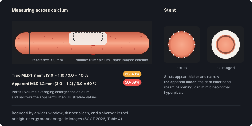
Partial volume averaging within each voxel blurs the interface between the lumen and calcium or a stent. The dense material appears larger and the stenosis is overestimated.
How to write it
Proximal LAD: calcified plaque with severe stenosis (70–99%), possibly overestimated due to calcium blooming.
Do not confuse: If the lumen can be seen through the calcium, report the highest visible grade with the caveat of possible overestimation; if blooming hides it, the segment is nonevaluable and the plaque is still described.
Source: SCCT 2026, Image quality, artifacts & pitfalls, e. Blooming artifact; Table 5, pp. 15 and 20 · “Blooming artifact results from partial volume averaging within a voxel, causing blurring at interfaces between coronary arteries and calcified plaques or stents and leading to overestimation of luminal stenosis”
Details
In practice. Review thin slices with a sharper kernel, or high-energy monoenergetic images if available. Over dense calcium, report the highest visible grade with the caveat of possible overestimation. A stent whose lumen cannot be distinguished is reported as nonevaluable for restenosis.
Pitfall. Measuring stenosis over dense calcium with the usual window exaggerates blooming. For that measurement, widen the window until the calcium edge is distinguishable.
Other sources. The SCCT 2023 standardized terminology prefers “partial volume averaging” and considers “calcium blooming” a legacy term that is not recommended. SCCT 2026 and CAD-RADS 2.0 keep “blooming”, the term used in this glossary. (SCCT 2023 terminology, Table 4)
In CCTA Map
With “Calcified” plaque from 50 to 99%, the template adds “grade possibly overestimated by calcium blooming”, including for plaque next to a stent in the same segment. In the impression, it suggests CT-FFR or functional testing if management depends on the grade; if the patient has a stent or graft, it suggests functional testing only, and if that territory already has a CT-FFR or CTP result, it does not repeat the suggestion. A stent marked “N” is written as “stent nonevaluable for restenosis due to artifact or small caliber”. Marking “N” sets the plaque aside rather than deleting it (a stent is kept): the segment is reported as nonevaluable and, with “Describe visible plaque”, the report names the calcified plaque without grading the stenosis. The plaque comes back when the N is removed; see “Non-interpretable (nondiagnostic) segment”.
Literal output of the map's template
Left anterior descending (LAD): proximal segment: stent nonevaluable for restenosis due to artifact or small caliber; the remainder is unremarkable. Branches D1, D2: no plaque.
Circumflex (LCx): proximal segment: calcified plaque causing minimal stenosis (1-24%); the rest of the vessel is unremarkable. Branches OM1, OM2, L-PLB: no plaque.
Right coronary (RCA): proximal segment: calcified plaque causing mild stenosis (25-49%); mid segment: calcified plaque producing minimal stenosis (1-24%); the rest of the vessel is unremarkable. Branches AM, R-PDA, R-PLB: no plaque.
Impression Nondiagnostic study for obstructive CAD. CAD-RADS N/P2/S.
Nonobstructive CAD in LCx and RCA.
CCTA has limited accuracy for in-stent restenosis in small-caliber stents (approximately <3 mm); consider functional imaging or invasive angiography if clinically suspected.
Nonevaluable segments: LAD proximal.
No numeric stenosis grade is assigned: CCTA cannot guide management in this setting and obstructive CAD cannot be excluded. Consider additional or alternative evaluation (repeat CCTA, functional/stress imaging, or invasive angiography).
Figure 1. Partial volume averaging spreads the attenuation of calcium and stent struts into neighboring voxels and narrows the apparent lumen. In this example, a true 40% stenosis measures 60% and moves from mild to moderate.
Also seen as:beam hardening artifact(RadLex name), beam hardening artefact, streak artifact(previous name for beam hardening not recommended by the SCCT 2023 terminology)not recommended
Metal, calcium and dense contrast preferentially absorb low-energy photons. Low-attenuation areas or streaks appear next to them and can mimic non-calcified plaque or a perfusion defect.
How to write it
Proximal RCA: calcified plaque with an adjacent low-attenuation band that decreases on high-energy monoenergetic images, consistent with beam-hardening artifact.
Do not confuse: Blooming enlarges calcium or stent struts and overestimates the stenosis; beam hardening leaves low-attenuation areas next to them that mimic non-calcified plaque or in-stent neointimal hyperplasia.
Source: SCCT 2026, Image quality, artifacts & pitfalls, d. Beam hardening and metallic artifacts; myocardium, pp. 15 and 19 · “Beam-hardening artifacts arise from preferential attenuation of low-energy photons by metal, calcium, or dense contrast, producing low-attenuation areas or streaks adjacent to bright artifacts”
Details
In practice. Suspect it when the hypoattenuation arises from calcium, metal or dense contrast. Review other phases, a higher level of iterative reconstruction or high-energy monoenergetic images. In the myocardium it is usually seen in the basal inferolateral wall and the apex.
Pitfall. Calling the low-attenuation band next to calcium a non-calcified component changes the plaque type. If it varies between phases or decreases on high-energy images, artifact is more likely.
Other sources. The SCCT 2023 standardized terminology discourages calling beam hardening a “streak artifact”. SCCT 2026 describes metallic artifact separately; it produces bright and dark streaks. (SCCT 2023 terminology, Table 4)
In CCTA Map
The map does not record this artifact. If the band turns out to be artifact, the plaque is marked “Calcified”, and the editor removes the “Low attenuation” and “Napkin-ring sign” features from that plaque and disables their buttons.
When photon counts are low, the image acquires a grainy texture with random bright and dark streaks. Excessive noise hides soft-tissue detail, especially non-calcified plaque.
How to write it
Excessive image noise related to body habitus, limiting detection of non-calcified plaque.
Source: SCCT 2026, Image quality, artifacts & pitfalls, a. Excessive noise; Table 4, pp. 14 and 15 · “Image noise from low photon counts appears as granular texture with random bright and dark streaks.”
Details
In practice. It is usually due to obesity, a tube current or tube voltage too low for the patient's size, or a phase reconstructed from the low-current portion of ECG-based tube current modulation. It is reduced with thicker slices, a smoother kernel or more iterative reconstruction, at the cost of spatial resolution.
Pitfall. Calling a noisy segment normal because no plaque is visible is an error, since noise hides non-calcified plaque. If plaque cannot be excluded, report the limitation.
Other sources. SCCT 2014 allowed noise to be measured as the standard deviation of HU in a region of homogeneous tissue. It also accepted reporting it as mild, moderate or severe, while acknowledging that these grades are not standardized. (SCCT 2014, section 10.6.1)
In CCTA Map
Noise has no dedicated control. It is expressed with the “Quality” selector in the “Draft report” panel and, if it prevents interpretation of a segment, that segment is marked with the “N” button in the vessel editor. With “nondiagnostic” and no segment marked “N”, the program applies the N rule to the whole study, as explained under “Overall image quality”. Marking “N” sets the segment’s plaque aside rather than deleting it, and it comes back when the N is removed; how to describe it in the report is explained under “Non-interpretable (nondiagnostic) segment”.
Also seen as:suboptimal opacification, poor contrast opacification, poor vessel opacification, inadequate contrast enhancement, insufficient luminal opacification
Enhancement is suboptimal when contrast opacifies the lumen poorly because of body size, slow circulation, injection technique or extravasation. Contrast mixing can mimic non-calcified plaque or stenosis.
How to write it
Distal LAD: nonevaluable due to suboptimal contrast enhancement.
Do not confuse: Contrast mixing changes between phases and is not accompanied by wall thickening; non-calcified plaque thickens the wall and persists in all phases.
Source: SCCT 2026, Image quality, artifacts & pitfalls, b. Suboptimal contrast enhancement, and g. False positive findings, pp. 14 and 17 · “Suboptimal contrast enhancement, contrast mixing, and flow artifacts can mimic non-calcified plaque or luminal stenosis”
Details
In practice. Review an equivocal area in other phases, with another window and on thicker slices to see whether the wall is thickened or only the lumen has low attenuation. Low-energy monoenergetic images increase luminal enhancement.
Pitfall. Reporting a poorly opacified distal segment as free of plaque. If the lumen cannot be distinguished, the segment is nonevaluable and is reported with its cause.
Other sources. The SCCT 2016 acquisition guideline considers intra-arterial opacification greater than 250 HU necessary for optimal images. SCCT 2026 sets no numeric threshold for calling enhancement suboptimal. (SCCT 2016 acquisition guideline, section 4.12.1)
In CCTA Map
Luminal opacification is not recorded separately. It is reflected in the “Quality” selector of the “Draft report” panel, and a segment that cannot be interpreted because of poor enhancement is marked with the “N” button in the vessel editor. With “nondiagnostic” and no segment marked “N”, the program applies the N rule to the whole study, as explained under “Overall image quality”. Marking “N” sets the segment’s plaque aside rather than deleting it, and it comes back when the N is removed; how to describe it in the report is explained under “Non-interpretable (nondiagnostic) segment”.
Multiplanar and curved multiplanar reformat MPR · CPR#
Also seen as:multiplanar reformation, multiplanar reconstruction, curved reconstruction(common usage; the SCCT 2023 terminology reserves “reconstruction” for generating images from the acquired projections), curved MPR(SCCT 2026 wording), CMPR(SCCT 2026 abbreviation; the SCCT 2023 terminology writes cMPR), curved planar reformat(RadLex name included in the SCCT 2023 terminology; source of the abbreviation CPR), curved multiplanar reconstruction(former term; the SCCT 2023 terminology recommends “curved multiplanar reformat”)not recommended, straightened multiplanar reformat, sMPR(a variant of the curved reformat that displays the vessel straight)
A multiplanar reformat (MPR) displays the acquired volume in any plane, and a curved reformat (CPR) follows the artery's centerline to display its entire course in a single longitudinal image, which can be rotated 360° around that axis. The cross-sectional view, or vessel short axis, is perpendicular to the centerline; it shows the lumen and the wall around the entire circumference and is usually the plane that best shows plaque size and composition.
How to write it
Each coronary artery was reviewed on axial images and on oblique, curved and short-axis multiplanar reformats.
Do not confuse: The axial image, which SCCT 2026 also calls “transverse”, is the body plane in which the acquisition is reconstructed; the vessel cross-section is perpendicular to the centerline and coincides with the axial plane only where the artery runs craniocaudally.
Source: SCCT 2026, image formats, b. Multiplanar reformats and c. Curved and straightened MPR, p. 2; coronary interpretation, p. 4; atherosclerotic plaque, p. 5; qualitative and quantitative stenosis grading, p. 7; false negatives, false positives and overestimation, pp. 15–17 · “Attention should be paid to post-processing artifacts including centerline misregistration, which can result in false-positive stenosis.”
Details
In practice. Stenosis is assessed on the cross-sectional view and in at least two mutually perpendicular longitudinal planes, and the lumen is compared with that of a normal reference segment. A centerline error can create a false stenosis, so before reporting a narrowing seen on the curved reformat, confirm that the centerline passes through the center of the lumen in that portion and that the narrowing persists on the cross-sectional view, in another plane and in the other reconstructed phases. The straightened reformat (sMPR) displays the vessel straight and helps to locate the most severe stenosis; quantification software measures diameters and areas on cross-sections taken from it or from a double-oblique orthogonal MPR. Coronal and sagittal planes help to recognize artifacts, such as the step of a misalignment artifact.
Pitfall. Grading stenosis on the automatic curved reformat alone. If the centerline strays from the center of the lumen, the reformat cuts the artery off its axis and can show a stenosis that does not exist; in addition, a single longitudinal plane can underestimate an eccentric stenosis.
Other sources. The SCCT 2023 standardized terminology recommends “multiplanar reformat” (MPR) and “curved multiplanar reformat” (cMPR), considers “curved multiplanar reconstruction” a former term that is not recommended, and reserves “reconstruction” for computing images from the acquired projections; the title of this entry uses the recommended terms, although “curved reconstruction” remains in common use. In the study by Anders 2011, cited by SCCT 2026, semiautomated vessel extraction with curved reformats did not improve accuracy over interactive MPR, and the authors do not recommend using automatic curved reformats alone, especially for less experienced readers. (SCCT 2023 terminology, Table 3; Anders 2011, Eur J Radiol, abstract)
CCTA image formats: what each one shows, what it is used for and its limitation.
Format
What it shows
Used for
Limitation or caution
Axial
The source images, with the highest spatial resolution and grayscale fidelity
Reviewing the entire volume; at 120 kVp, the window starts at a level of 300 and a width of 800 and is adjusted to body habitus, calcium and contrast enhancement
A tortuous vessel is hard to follow
Oblique and cross-sectional MPR
The volume in any plane, at the thinnest available slice thickness (for example, 0.6 or 0.4 mm)
Following the vessel and seeing the plaque and its effect on the lumen and the wall
Stenosis is confirmed on the cross-sectional view and in two perpendicular longitudinal planes
Curved (CPR)
The entire course of the vessel in one longitudinal image that rotates 360°
Following tortuous vessels and obtaining longitudinal and cross-sectional views at any point
A centerline error creates false stenoses, and a single plane can underestimate an eccentric stenosis
Straightened (sMPR)
The vessel displayed as a straight line
Locating the most severe stenosis, assessing plaque and pericoronary fat, and quantification
It is derived from the curved reformat, so its centerline is checked as well
MIP
The highest-attenuation voxel along each line of sight: the contrast-enhanced lumen and calcium
With 5 to 10 mm slabs, origin, course and lesion location; with thin slabs, lesions that are easily missed
Not for grading: it underestimates non-calcified plaque, overestimates calcified plaque and hides perfusion defects
MinIP
The lowest-attenuation voxel
Myocardial perfusion defects and valves
SCCT 2026 mentions it only for the myocardium and the valves
It does not quantify stenosis, does not assess calcified plaque well and does not show non-calcified plaque
Source: SCCT 2026, “CCTA image formats”, items a–e and h, pp. 2–3; quantitative grading, p. 7; false negatives, p. 15; myocardium, p. 19
In CCTA Map
The map does not display images or reformats: it records what the radiologist has already assessed on the workstation, and it does not record the plane in which each stenosis was graded. The grade chosen with the 0–5 “Severity” buttons or with the “Stenosis grade” slider should be the one confirmed on the cross-sectional view and in two longitudinal planes.
Maximum and minimum intensity projection MIP · MinIP#
Also seen as:maximum intensity projection, minimum intensity projection, thin-slab MIP(a slab a few millimeters thick), MiniP(spelling of the abbreviation in the myocardium section of SCCT 2026)
A maximum intensity projection (MIP) displays, for each line of sight through a slab of the chosen thickness, the voxel with the highest attenuation, and a minimum intensity projection (MinIP) displays the voxel with the lowest attenuation. MIP highlights the contrast-enhanced lumen and calcium; MinIP highlights hypodense tissue, such as a myocardial perfusion defect. MIP loses attenuation information in depth and is not reliable for grading stenosis.
How to write it
Subendocardial hypoattenuating area in the mid inferior segment of the left ventricle, seen on short-axis MinIP reformats with a narrow window.
This example assumes: The hypoattenuation persists in several phases of the cardiac cycle; if it appears, disappears or moves between phases, it suggests artifact, such as beam hardening.
Do not confuse: At the same slab thickness, MIP shows the densest voxel and masks a perfusion defect, MinIP shows the least dense voxel and highlights it, and averaged MPR shows the mean attenuation.
Source: SCCT 2026, image formats, d. Maximal intensity projection and h. Other image types, p. 3; false negatives and false positives, pp. 15–16; myocardium, p. 19. SCCT 2023 terminology, Table 3 · “MIP should not be interpreted in isolation because it lacks in-depth attenuation information and is unreliable for stenosis assessment, underestimating noncalcified plaque and overestimating calcified disease”
Details
In practice. SCCT 2026 suggests MIP slabs of 5 to 10 mm to follow long segments with little noise and to show the origin, course and location of lesions. Thin-slab MIP helps to find lesions that are easily missed near bifurcations, in small vessels and distal to a graft anastomosis, and to clarify an apparent stenosis in a tortuous or kinked vessel. What is seen on MIP is then graded on thin-slice MPR. In the myocardium, perfusion is best assessed in several planes (short axis, four-chamber and two-chamber), with 5 to 8 mm averaged MPR or MinIP and a narrow window, for example a width of 200 and a level of 100 HU; MinIP is also used to assess the valves.
Pitfall. Grading a stenosis on MIP or looking for perfusion defects on it. SCCT 2026 warns that MIP underestimates non-calcified plaque, overestimates calcified plaque and should not be interpreted in isolation, and that it should be avoided in the myocardium because it masks defects.
Other sources. Karlo 2012, a review that SCCT 2026 cites for false-negative and false-positive findings, describes MIP as an angiography-like image, uses 3 to 5 mm slabs to locate plaques and advises against slabs thicker than 10 mm, which can hide lesions or create false positives from wall calcium or nearby stents. In SCCT 2021, first-pass perfusion is reviewed with 8 mm short-axis MinIP and a narrow window, and late enhancement, in which scar retains iodine and appears denser, with 5 or 10 mm thick MPR or with MIP. (Karlo 2012, Insights Imaging, “Post-processing” and “Evaluation of coronary arteries: step one”; SCCT 2021, section 4.8, p. 208)
In CCTA Map
The map does not display images or record which reformat showed a finding. A hypoattenuating area of the myocardium on resting CCTA has no field of its own: it is described with “✎ Edit draft”, and the edited draft no longer updates from the map. The “Ischemia (I)” section of “Modifiers P · I ▾” records only the result of CT-FFR or of stress CT myocardial perfusion, under the “CT-FFR” and “CTP” pathways.
Self-assessment
Short questions to check the key points. The answer appears once you choose an option.
1The mid RCA remains blurred by motion after all reconstructed phases have been reviewed, and the rest of the coronary tree is interpretable. According to SCCT 2026, what should be stated in the report?
The guideline asks you to name the segments that could not be interpreted and the responsible artifact. This tells the referring physician which part of the tree was not assessed and why. See the term
2On the curved MPR, the distal LAD shows a narrowing with straight edges. On the sagittal plane at that level, a horizontal line offsets the cardiac contour, but the sternum and spine appear continuous. What is the most likely explanation?
A step at a slab boundary that spares the sternum and spine points to rhythm-related misalignment. Respiratory motion would displace those structures as well. Confirm by reconstructing another phase. See the term
3A densely calcified plaque in the proximal LAD produces a stenosis that still measures about 75% with a wide window and a sharper kernel. How should it be reported according to SCCT 2026?
Table 5 recommends reporting the highest visible grade over dense calcium with the caveat of possible overestimation from blooming. Some readers subtract 10 to 20%, but doing so without saying so hides the uncertainty from the referring physician. See the term
4A low-attenuation band lies next to a dense calcification in the proximal RCA. It changes shape in another phase and almost disappears on the high-energy monoenergetic reconstruction. What does it represent?
Beam hardening produces low-attenuation areas next to calcium that mimic non-calcified plaque. It improves in other phases and on high-energy images, whereas true plaque persists. See the term
5In a patient with a BMI of 45, the study has a grainy texture that makes the arterial wall hard to see. Which reconstruction change reduces noise?
Thicker slices, a smoother kernel and a higher level of iterative reconstruction reduce noise at the cost of spatial resolution. Thin slices and a sharp kernel increase it. See the term
Rajiah PS, Alkadhi H, Andreini D, Bullock-Palmer RP, Chinnaiyan K, Chow B, et al. Interpretation and reporting of coronary computed tomographic angiography (2026 update): an expert consensus document of the Society of Cardiovascular Computed Tomography (SCCT). J Cardiovasc Comput Tomogr. 2026. doi:10.1016/j.jcct.2026.08.014 Version of record published online in September 2026 under CC BY 4.0, not yet assigned to an issue; cited pages refer to the PDF of this published version.
Cury RC, Leipsic J, Abbara S, Achenbach S, Berman D, Bittencourt M, et al. CAD-RADS 2.0 - 2022 Coronary Artery Disease-Reporting and Data System: an expert consensus document of the SCCT, ACC, ACR and NASCI. J Cardiovasc Comput Tomogr. 2022;16(6):536-57. doi:10.1016/j.jcct.2022.07.002
Koweek L, Achenbach S, Berman DS, Carr JJ, Cury RC, Ghoshhajra B, et al. Standardized medical terminology for cardiac computed tomography 2023 update: an expert consensus document of the SCCT, AAPM, ACR, NASCI and RSNA. J Cardiovasc Comput Tomogr. 2023;17(5):345-54. doi:10.1016/j.jcct.2023.06.002
Narula J, Chandrashekhar Y, Ahmadi A, Abbara S, Berman DS, Blankstein R, et al. SCCT 2021 expert consensus document on coronary computed tomographic angiography: a report of the Society of Cardiovascular Computed Tomography. J Cardiovasc Comput Tomogr. 2021;15(3):192-217. doi:10.1016/j.jcct.2020.11.001
Neves PO, Andrade J, Monção H. Coronary artery calcium score: current status. Radiol Bras. 2017;50(3):182-9.
Tamburino C, Tomasello SD, Capodanno D, Di Salvo ME, Marzà F, Galassi AR. Long-term follow-up after drug eluting stent implantation in left main trifurcations. EuroIntervention. 2009;5(4):432-7. doi:10.4244/eijv5i4a68
Kovacevic M, Burzotta F, Elharty S, Besis G, Aurigemma C, Romagnoli E, et al. Left main trifurcation and its percutaneous treatment: what is known so far? Circ Cardiovasc Interv. 2021;14(3):e009872. doi:10.1161/CIRCINTERVENTIONS.120.009872
Kim C, Park CH, Lee BY, et al. 2024 consensus statement on coronary stenosis and plaque evaluation in CT angiography from the Asian Society of Cardiovascular Imaging-Practical Tutorial (ASCI-PT). Korean J Radiol. 2024;25(4):331-42. doi:10.3348/kjr.2024.0112
Sources for the “Other sources” notes
Leipsic J, Abbara S, Achenbach S, Cury R, Earls JP, Mancini GBJ, et al. SCCT guidelines for the interpretation and reporting of coronary CT angiography: a report of the Society of Cardiovascular Computed Tomography Guidelines Committee. J Cardiovasc Comput Tomogr. 2014;8(5):342-58. doi:10.1016/j.jcct.2014.07.003
Cury RC, Abbara S, Achenbach S, et al. CAD-RADS: Coronary Artery Disease – Reporting and Data System. J Cardiovasc Comput Tomogr. 2016;10(4):269-81. doi:10.1016/j.jcct.2016.04.005
Nieman K, García-García HM, Hideo-Kajita A, et al. Standards for quantitative assessments by coronary computed tomography angiography (CCTA): an expert consensus document of the SCCT. J Cardiovasc Comput Tomogr. 2024;18(5):429-43. doi:10.1016/j.jcct.2024.05.232
Koweek L, Achenbach S, Berman DS, et al. Standardized medical terminology for cardiac computed tomography 2023 update: an expert consensus document of the Society of Cardiovascular Computed Tomography (SCCT), American Association of Physicists in Medicine (AAPM), American College of Radiology (ACR), North American Society for Cardiovascular Imaging (NASCI), and Radiological Society of North America (RSNA). Radiol Cardiothorac Imaging. 2023;5(4):e230167. doi:10.1148/ryct.230167
Abbara S, Blanke P, Maroules CD, et al. SCCT guidelines for the performance and acquisition of coronary computed tomographic angiography: a report of the Society of Cardiovascular Computed Tomography Guidelines Committee. Endorsed by the North American Society for Cardiovascular Imaging (NASCI). J Cardiovasc Comput Tomogr. 2016;10(6):435-49. doi:10.1016/j.jcct.2016.10.002
This glossary translates and adapts definitions, tables and figures from the SCCT 2026 consensus: Rajiah PS, et al. Interpretation and reporting of coronary computed tomographic angiography (2026 update): an expert consensus document of the Society of Cardiovascular Computed Tomography (SCCT). J Cardiovasc Comput Tomogr. 2026. doi:10.1016/j.jcct.2026.08.014, published under the CC BY 4.0 license. Changes: plain-language rewording and reorganized tables; the diagrams are dynamic schematics of the map and do not reproduce the guideline figures exactly. The SCCT has not reviewed or endorsed this glossary. CC BY 4.0